Wednesday, October 14, 2015

The Rise of Multidrug-Resistant Strains and Need for New Therapeutic Approaches

Cara N. Wilder, Ph.D.

The discovery of antibiotics in the early twentieth century has revolutionized the treatment of infectious diseases, saving millions of lives and easing the suffering of many. However, as the structure and function of antibiotics has evolved through the efforts of biotech and pharmaceutical companies, microorganisms have evolved in parallel, fashioning novel and effective methods to avoid therapeutic biocide. In the last several decades, this concern has become more pronounced with the emergence of multidrug-resistant organisms in both community- and hospital-acquired infections, resulting in increased morbidity, mortality, and health-care expense. Here, we will discuss the growing threat of antimicrobial resistance, novel therapeutic approaches, and the importance of antimicrobial-resistant reference strains in reducing the emergence and spread of multidrug-resistant infections.

Antimicrobial resistance is currently recognized as one of the greatest threats to human health worldwide1. According to the World Health Organization (WHO), antimicrobial-resistant strains are present in all parts of the world, and new resistance mechanisms continue to emerge and spread globally2. In the United States alone, at least 2 million people become infected with antibiotic-resistant bacteria, with approximately 23,000 associated deaths each year3. These patients are typically at an increased risk of more debilitating clinical outcomes and death, and consume more healthcare resources as compared to patients infected with a drug-sensitive strain of the same species2. Infection with these microorganisms is further complicated by the limited number of effective therapeutic options. In some cases, clinicians have resorted to using older drugs, for which there may be limited data on safety and efficacy to guide the dosage or duration4,5. Overall, this highlights the dire need for novel and effective therapeutics to treat multidrug-resistant infections.

As the number of antibiotics effective against multidrug-resistant strains is beginning to dwindle, there have been a variety of efforts made to evaluate novel therapeutic options. For example, one novel mechanism used in a 2010 study by Nicolosi et al. employed the use of fusogenic liposomes to transport vancomycin into Gram-negative cells6. Under normal conditions, vancomycin is only used to treat Gram-positive organisms. It is inactive against Gram-negative bacteria due to the different mechanism in which Gram-negative bacteria produce their cell walls, as well as other factors relating to entering the outer membrane of Gram-negative organisms. Fusogenic liposomes offer an alternative method for the delivery of the antibiotic across the gram-negative outer membrane. These small unilamellar liposome vesicles are able to adhere to and fuse with the external membrane; thus, if you load these liposome vesicles with an antibiotic such as vancomycin, you can transport the antibiotic across the membrane. Overall, this offers a promising mechanism for extending the use of current antibiotics.

Another emerging therapeutic approach against multidrug-resistant strains is the use of antimicrobial peptides. For example, in a 2010 study by Routsias et al., the group found that human beta-defensin 2, which is a naturally occurring peptide, exhibited high bactericidal activity against Acinetobacter baumannii. As antimicrobial peptides are naturally occurring, are bactericidal, and are broad-spectrum, they offer a promising mechanism for treating drug-resistant infections7. Antisense agents also provide another potential therapeutic approach for treating drug-resistant infections by inhibiting resistance mechanisms directly at the nucleic acid level. These oligonucleotides could potentially be designed to bind specific mRNA or DNA sequences that confer resistance to antibiotics, thus blocking either translation or gene transcription, rendering the cell susceptible to antibiotic treatment. However, as these nucleic acids would have no intrinsic antibacterial activity of their own, they would have to be used in conjunction with antibiotics as needed8.

Bacteriophage T4. Photo credit: David
Gregory and Debbie Marshall
Before the use of antibiotics, some efforts were made to use bacteriophages for the treatment of bacterial infections. Following the first isolation of a bacteriophage in 1917, bacteriophages have been extensively used and developed in several countries throughout the former Soviet Union9. The primary advantage to this form of treatment is the specificity of a bacteriophage toward a specific bacterial target. This would allow for direct killing of a bacterial pathogen without harming the resident microflora or the patient. In turn, this would help reduce the likelihood of opportunistic, secondary infections from occurring. However, this specificity also poses a potential problem in that to effectively treat an infection, the causative agent must be properly diagnosed10,11. Further, it is possible for bacterial strains to become resistant to certain types of bacteriophages following continual exposure; however, this could be potentially overcome through the use of genetically modified bacteriophages.

Lastly, there are some efforts being made to help support the development of new antibiotics. In 2010, the Infectious Diseases Society of America (IDSA) launched a collaboration entitled the “10 x ‘20” initiative to help create a global antibacterial drug research and development enterprise with the power to develop 10 new, safe, and effective antibiotics by the year 202012. Here, the IDSA is supporting the development of 10 new systemic antibacterial drugs through the development of new drugs from existing antibiotic classes or through the discovery of novel antibiotic classes not previously known12. To date, the initiative has spurred the development of two novel antibiotics, which were recently approved in the United States by the Food and Drug Administration (FDA)13,14.

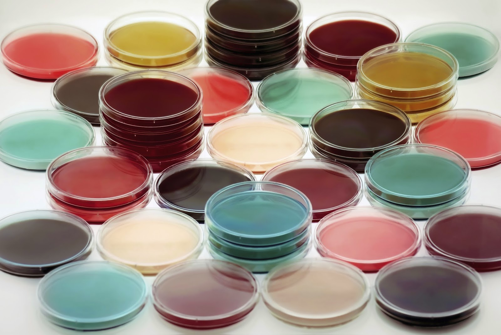
Regardless of the type of therapeutic approach, it is important to ensure that the novel treatment is both safe for patient use and effective against a variety of drug-resistant strains. This can be evaluated through in vitro analyses using authenticated strains and cell lines. Characterized microbial strains with known antibiotic susceptibility profiles are ideal for evaluating the efficacy of a novel therapeutic against different species that vary in their resistance/susceptibility profiles. These strains would also be effective in the development and validation of novel detection methods as it would allow a mechanism to evaluate the sensitivity and specificity against various species and mechanisms of drug resistance. For analyzing the safety of the therapeutic, primary cells derived from various organ systems and donors can provide a quick mechanism for screening therapeutics for any potential toxic effects.

To support this need, ATCC offers a complete set of solutions to advance multidrug-resistance research, including drug-resistant microbial reference strains from various clinical and environmental sources, primary cells for drug toxicity screening studies, as well as media and reagents that support growth. Each of these reference materials are produced under ISO 9001:2008 certified and ISO/IEC 17025:2005 accredited processes, ensuring reliability and reproducibility of research data. Further, to ensure that each of these products are of the highest quality before they reach your laboratory, ATCC has authenticated each strain using phenotypic and genotypic analyses to guarantee species identification, culture purity, and biochemical consistency.

Overall, multidrug-resistant microbial strains are continually implicated in a number of infections worldwide, resulting in significant increases in morbidity, mortality, and health care expense. To help control the emergence and spread of these pathogens, discovering new and effective treatment methods is of the upmost importance. To support this need, ATCC offers fully authenticated, characterized strains and cell lines for use as positive controls. These cultures are ideal for evaluating the efficacy and toxicity of novel therapeutic treatments. In conclusion, microorganisms are rapidly evolving or acquiring mechanisms of resistance to antibiotics. Only through aggressive action and scientific ingenuity can the emergence and spread of multidrug resistance be prevented.


References
  1. Walker, B. et al. Environment. Looming global-scale failures and missing institutions. Science 325, 1345-1346, doi:10.1126/science.1175325 (2009).
  2. WHO. Fact sheet N°194 - Antimicrobial resistance, <http://www.who.int/mediacentre/factsheets/fs194/en/> (2014).
  3. CDC. Antibiotic/Antimicrobial Resistance, <http://www.cdc.gov/drugresistance/> (2014).
  4. Boucher, H. W. et al. Bad bugs, no drugs: no ESKAPE! An update from the Infectious Diseases Society of America. Clinical infectious diseases : an official publication of the Infectious Diseases Society of America 48, 1-12, doi:10.1086/595011 (2009).
  5. Cassir, N., Rolain, J. M. & Brouqui, P. A new strategy to fight antimicrobial resistance: the revival of old antibiotics. Frontiers in microbiology 5, 551, doi:10.3389/fmicb.2014.00551 (2014).
  6. Nicolosi, D., Scalia, M., Nicolosi, V. M. & Pignatello, R. Encapsulation in fusogenic liposomes broadens the spectrum of action of vancomycin against Gram-negative bacteria. International journal of antimicrobial agents 35, 553-558, doi:10.1016/j.ijantimicag.2010.01.015 (2010).
  7. Routsias, J. G., Karagounis, P., Parvulesku, G., Legakis, N. J. & Tsakris, A. In vitro bactericidal activity of human beta-defensin 2 against nosocomial strains. Peptides 31, 1654-1660, doi:10.1016/j.peptides.2010.06.010 (2010).
  8. Woodford, N., Wareham, D. W. & Group, U. K. A. A. S. Tackling antibiotic resistance: a dose of common antisense? The Journal of antimicrobial chemotherapy 63, 225-229, doi:10.1093/jac/dkn467 (2009).
  9. Mandal, S. M. et al. Challenges and future prospects of antibiotic therapy: from peptides to phages utilization. Frontiers in pharmacology 5, 105, doi:10.3389/fphar.2014.00105 (2014).
  10. Bragg, R., van der Westhuizen, W., Lee, J. Y., Coetsee, E. & Boucher, C. Bacteriophages as potential treatment option for antibiotic resistant bacteria. Advances in experimental medicine and biology 807, 97-110, doi:10.1007/978-81-322-1777-0_7 (2014).
  11. Tiwari, R., Dhama, K., Kumar, A., Rahal, A. & Kapoor, S. Bacteriophage therapy for safeguarding animal and human health: a review. Pakistan journal of biological sciences : PJBS 17, 301-315 (2014).
  12. ISDA. The 10 × '20 Initiative: Pursuing a Global Commitment to Develop 10 New Antibacterial Drugs by 2020. Clinical infectious diseases : an official publication of the Infectious Diseases Society of America 50, 1081-1083 (2010).
  13. Boucher, H. W. et al. 10 x '20 Progress--development of new drugs active against gram-negative bacilli: an update from the Infectious Diseases Society of America. Clinical infectious diseases : an official publication of the Infectious Diseases Society of America 56, 1685-1694, doi:10.1093/cid/cit152 (2013).
  14. Murray, B. Progress in 10 x '20 Initiative Shows Promise but More Action Needed. Infection Control Today (June 23, 2014).


Thursday, September 3, 2015

The role of microorganisms in the induction of cancer

Cara N. Wilder, Ph.D.

Since the beginning of the 20th century, microorganisms have been known to play a significant role in the development of cancer in animals1. However, our understanding on the extent of this issue in humans is still continuing to expand. Currently, it is estimated that microbial infections are linked to approximately 15-20% of human cancers throughout the world1-3. This presents an enormous potential for the prevention of cancer through the control of infectious disease.

In a 2006 International Journal of Cancer study, it was estimated that the total number of cancers caused by microbial infection in 2002 was approximately 17.8% of the global cancer burden2. Of this subset, most of the burden was attributable to viral infections (12.1%), followed by infection with Helicobacter pylori (5.6%), with a small proportion due to parasitic infection (0.1%) (Table 1)2. Further, the percentage of these infection-attributable cancers was higher in developing countries as compared to developed countries, reflecting the higher prevalence of these organisms, and perhaps the need for more readily-available healthcare.

Table 1. Examples of microbial-associated cancers2,4
Agent
Type
Cancer
Epstein-Barr virus (EBV)
Herpesvirus
Hodgkin’s lymphoma, Burkitt’s lymphoma, Nasopharyngeal carcinoma
Helicobacter pylori
Bacterium
Gastric adenocarcinoma
Hepatitis B virus (HBV)
Hepadnavirus
Hepatocellular carcinoma
Hepatitis C virus (HCV)
Flavivirus
Hepatocellular carcinoma
Human herpesvirus 8 (HHV-8)
Herpesvirus
Kaposi’s sarcoma
Human papillomavirus (HPV)
Papillomavirus
Anogenital carcinoma, Oropharyngeal carcinoma
Human T-lymphotropic virus 1 (HTLV-1)
Retrovirus
Adult T-cell lymphoma
Opisthorchis viverrini
Trematode
Cholangiocarcinoma
Schistosoma haematobium
Trematode
Bladder squamous cell carcinoma

The ability of these microorganisms to cause cancer greatly depends on the context of the host-microbial relationship. These interactions can vary significantly due to a number of factors such as the oncogenic potential of the strain, how the strain interacts with the host’s cells and immune system, if the infection causes long-term inflammation, host genotype and phenotype, environmental factors, microbial load, and the composition of the host’s microbiome1,4. Further, there is some speculation that the ability for certain cancers to run within a family may in part be due to the intra-familial transmission of oncogenic microorganisms. In this latter case, indigenous organisms acquired from a family member may be preadapted to the next host, which could modify the risk of disease5,6. Regardless of the mechanism, it is clear that preventing or controlling microbial infections may help reduce the number of cancer cases throughout the world.

There are a number of methods that can be used to help prevent cancers associated with microbial infection. Taking measures to avoid exposure to strains associated with infection-attributable cancers, such as circumventing the habitats of harmful species or abstaining from activities that promote transmission, can help reduce the likelihood of infection. Another approach is through the use of vaccination. Currently, vaccines are available against the hepatitis B virus as well as human papillomavirus types 16 and 18. As these vaccines have already demonstrated efficacy in preventing infection, they have the potential to significantly reduce healthcare cost as well as the incidence of liver and anogenital cancer, respectively. If vaccination is not possible, available antimicrobials can be used to help mediate or cure an established infection. For example, in the treatment of H. pylori, the administration of triple therapy regimens for 10-14 days consisting of a proton pump inhibitor, amoxicillin, and clarithromycin are typically recommended, and have reported cure rates from 85-90%7.

Overall, infection-attributable cancers represent a significant, albeit preventable, proportion of the global cancer burden. There are currently a number of therapeutic options to help stave off, control, or treat many known oncogenic infections; however, more research is still needed to expand the range of effective therapeutic options as well as help identify additional microbial strains that have the potential to cause cancer. To that end, a number of reputable biological resource centers, such as ATCC, offer a variety of microorganisms, nucleic acids, and cell lines that can be used to help support the research in this field. Only through hard work and dedicated research can we help significantly reduce the number of cancer cases throughout the world.


References
  1. American Cancer Society. Infections that can lead to cancer, <http://www.cancer.org/cancer/cancercauses/othercarcinogens/infectiousagents/infectiousagentsandcancer/infectious-agents-and-cancer-intro> (2014).
  2. Parkin, D. M. The global health burden of infection-associated cancers in the year 2002. International journal of cancer. Journal international du cancer 118, 3030-3044, doi:10.1002/ijc.21731 (2006).
  3. WHO. Global status report on non communicable disease 2010. WHO Library Cataloguing-in-Publication Data (2011).
  4. Blaser, M. J. Understanding Microbe-Induced Cancers. Cancer Prevention Research 1, 15-20 (2008).
  5. Blaser, M. J. & Kirschner, D. The equilibria that allow bacterial persistence in human hosts. Nature 449, 843-849, doi:10.1038/nature06198 (2007).
  6. Blaser, M. J., Nomura, A., Lee, J., Stemmerman, G. N. & Perez-Perez, G. I. Early-life family structure and microbially induced cancer risk. PLoS medicine 4, e7, doi:10.1371/journal.pmed.0040007 (2007).
  7. Santacroce, L. & Bhutani, M. Helicobacter Pylori Infection Treatment & Management, <http://emedicine.medscape.com/article/176938-treatment> (2014).


Monday, February 23, 2015

Using Next-Generation Sequencing for Infectious Disease Research – Understanding Microbial Pathogens from Within

Cara N. Wilder, Ph.D.

Ever since James Watson and Francis Crick derived the double-helix model for the structure of DNA in 1953, the field of genomics has exponentially burgeoned into a vast array of molecular technologies. Of the current assortment of available genomic tools, next-generation sequencing (NGS) has proven to be of great value, particularly in the field of infectious disease. Here, the knowledge of DNA and RNA sequences has become indispensable for a variety of applications, including microbial identification and detection, evaluating the evolution and regulation of virulence, identifying drug resistance markers, detecting antigenic targets for vaccine development, and delineating community outbreaks.

NGS technologies are typically described as non-Sanger-based high-throughput DNA sequencing platforms that enable the analysis of millions of DNA fragments in parallel1. This ability to multiplex provides unprecedented scalability and has contributed to a gradual reduction in reagent costs, allowing whole-genome sequencing (WGS) to become accessible and practical to researchers around the world. Over the last decade, a number of platforms and methods have been developed that enable de novo sequencing, re-sequencing, transcriptome profiling, and cDNA sequencing.

In the field of infectious disease, NGS affords a means for reviewing the complete genetic make-up of microbial pathogens through de novo sequencing or re-sequencing, regardless of the fastidious or non-fastidious nature of the strain. This ability to review an entire genome, or individual genes or operons, provides a unique opportunity for clinical diagnostics, the identification of potential antigenic targets for vaccine development, and epidemiological surveillance. Currently, the diagnosis of microbial infections typically begins with patient observation and the collection of specimens for microbiological processing for various diagnostic tests. Depending on the causative agent, these diagnostics tests may include serology and analyzing specimens for the presence of immune cells via microscopy. For some bacterial infections, culture-based growth techniques, matrix-assisted laser desorption/ionization time-of-flight mass spectrometry (MALDI-TOF MS), and polymerase chain reaction (PCR) are also used for microbial identification and analysis. If employed, NGS could potentially be used to identify a pathogen without prior knowledge of the target, as well as discern the presence of any virulence determinants or genes conferring drug resistance2-4. In turn, this would provide an additional clinical tool that may help patients receive a more precise diagnosis and efficient treatment regimen.

In addition to clinical diagnostics, NGS offers a potential tool for vaccine development and evaluation. For example, NGS can allow for the surveillance of pathogen evolution. For rapidly mutating pathogens such as the influenza virus, this could provide an opportunity to help identify antigenic drift within the microbial population and use that knowledge to create a better, more specific vaccine. NGS could also be used for reverse vaccinology, where pathogen sequences are used to identify putative surface antigens that could potentially be targeted for the development of a novel vaccine preparation4. In accordance with this latter application, the expression of the putative surface antigens can also be examined through the evaluation of RNA sequences during different stages throughout the pathogen life cycle. As some strains require more than one species for survival and replication, analyzing RNA expression can help ensure that the vaccine target is actively expressed in the human host to ensure an efficient immunological response. Lastly, NGS could be applied in vaccine safety analyses by detecting the presence of viral contaminants in cell culture used in vaccine development or identifying virulent mutations in live-attenuated vaccines prepared with highly pathogenic, genetically unstable strains prior to the use of the vaccine5,6. Overall, NGS could help contribute to the development of safer vaccines that provide a more effective immune response.

Lastly, NGS is gradually being employed in the epidemiological analysis of cultures for outbreak detection and infection control. Using genomics, outbreak strains have been identified at the species level and below with extraordinarily high resolution. Through the analysis of these strains, population genetic studies can contribute to a better understanding of important genetic markers among strains while also tracking transmission events during outbreaks. In turn, this allows for the development of a tailored control program to help limit the further spread of disease7. A recent example of the use of NGS in epidemiological surveillance was during the analysis of a 2011 hospital outbreak of methicillin-resistant Staphylococcus aureus (MRSA)8. Here, WGS was used to identify hospitalized infant patients infected with MRSA and determine the source of transmission. Through this study, 26 related cases of MRSA were identified and it was demonstrated that transmission occurred within the hospital between mother and patient, among mothers on a postnatal ward, and in the community. Moreover, WGS confirmed that MRSA carriage by a staff member allowed the outbreak to persist during periods of no known infection. Overall, this study demonstrated that NGS technology enabled a more precise identification of patients involved in an outbreak, allowing for the implementation of an effective infection control program that helped halt the spread of disease.

Despite the numerous advantages of NGS in the analysis of infectious diseases, there are a few challenges that may need to be overcome. For example, for NGS data to be reliable, particularly with regard to re-sequencing and microbial identification, it is important that validated sequence data is readily accessible. Well-renowned sequence databases (e.g. GenBank) provide an annotated collection of publically available genome sequences from authenticated type strains, such as those obtained from prominent biological resource centers like ATCC. As type strains are the nomenclatural origin of a species or subspecies, they are typically fully authenticated and exhibit all of the relevant phenotypic and genotypic properties described in original taxonomic circumscriptions, making them ideal reference standards for NGS. Extensive bioinformatics to analyze sequences from assembly to annotation, which necessitates expertise and time, could pose another challenge for NGS clinical use. This can be potentially circumvented through the development of automated tools for sequence analysis combined with the assemblage of exhaustive databases containing all known genome sequences3Lastly, there is the possibility that host DNA will be analyzed alongside microbial nucleic acids, which may make it difficult to discern the target genomics. To overcome this, the microbial to host nucleic acid ratio must be increased through procedures such as hydrolysis or chemical treatment4.

In conclusion, clinical microbiology laboratories face a number of challenges regarding the diagnosis and effective treatment of infectious diseases. NGS could provide an additional insight to aid in clinical diagnostics while offering an effective mechanism for the identification of potential vaccine targets and a way to track outbreaks. Only through continual efforts in the advancement of this technology can NGS become fully ingrained in the field of infectious disease as a routine diagnostic tool.


References
  1. Nature. Next-generation sequencing – Definition. http://www.nature.com/subjects/next-generation-sequencing, 2015. 
  2. Wain J, Mavrogiorgou E. Next-generation sequencing in clinical microbiology. Expert Rev Mol Diagn 13(3): 225-227, 2013. 
  3. Fournier PE, et al. Modern clinical microbiology: new challenges and solutions. Nat Rev Microbiol 11: 574-585, 2013. 
  4. Lecuit M, Eloit M. The diagnosis of infectious disease by whole genome next generation sequence: a new era is opening. Front Cell Infect Microbiol 4: 25, 2014. 
  5. Luciani F, et al. Next generation deep sequencing and vaccine design: today and tomorrow. Trends in Biotechnology 30(9): 443-452, 2012. 
  6. Neverov A, Chumakov K. Massively parallel sequencing for monitoring genetic consistency and quality control of live viral vaccines. Proc Natl Acad Sci 107(46): 20063-20068, 2010. 
  7. Tang P, Gardy JL. Stopping outbreaks with real-time genomic epidemiology. Genome Med 6(11): 104, 2014. 
  8. Harris SR, et al. Whole-genome sequencing for analysis of an outbreak of methicillin-resistant Staphylococcus aureus: a descriptive study. Lancet Infect Dis 13(2): 130-136, 2013.

Tuesday, September 23, 2014

Current techniques used in the quality control of culture media required for pharmaceutical microbiology

Cara N. Wilder, Ph.D.

Culture media is a basic tool of microbiology, supporting most microbiological assays such as propagation, obtaining pure cultures, enumerating cells, preservation, and selecting microorganisms. For pharmaceutical quality control laboratories, media is frequently used for environmental monitoring, sterility testing, and microbial enumeration tests. As such, the quality of microbial-based tests greatly depends on the quality of the culture media; if the media is not the right quality, it undermines all of the tests that the media is used for. Therefore, safeguarding the quality of culture media through routine quality control testing can help ensure reliable, consistent microbiological test results.

Culture media are traditionally defined as substances that encourage the growth, support, and survival of microorganisms. This is achieved through the preparation of liquid broth or solid agar media comprising reagents required to support microbial growth, including basic nutrients, energy sources, growth factors, minerals, buffer salts, and metals1. To help minimize lot-to-lot variability, culture media manufacturers often attempt to standardize the preparation of media; however, there are frequently unavoidable differences in raw materials from natural sources, intra-lab skill level, or the storage and shipping conditions of the media. To help counter this, culture media should be physically inspected for color, clarity, damage, pH, and gel strength following all preparation steps. Moreover, media that has not been quality control tested or assessed should be quarantined to prevent premature use.

In addition to the physical inspection of culture media, it is important to analyze media for microbiological characteristics. As these media are frequently used for the analysis of sterility throughout the manufacturing process, or for the detection of objectionable microorganisms, it is imperative that the media is not only sterile, but able to promote the growth of any possible contaminants. For the analysis of media sterility, uninoculated media is commonly incubated. Here, the growth temperature and incubation period will depend on the type of media being analyzed. Media exhibiting the absence of growth following the recommended incubation period are considered sterile. In contrast, growth promotion testing is one of the most important quality control tests performed on media and is used to determine if the media in question is able to promote and sustain growth. The primary objective of the test is to detect if a new batch of media is functional and of the same standard as the most recent batch of media tested, as well as ensures the consistent use of standardized media between labs.

Currently, there are several approaches that can be taken for testing media for growth promotion. For agar-based media, a simple method of analysis is to serially dilute microbial strains and plate them on test media using the spread plate technique. These agar plates are then compared to the growth characteristics of a control plate, which is a batch of media that has been previously assessed for growth promotion and approved for use. Two other, more robust, approaches include the Miles-Misra and the ecometric techniques2. The Miles-Misra method is a quantitative technique used to determine the surface viable count. Here, droplets of titered microbial suspensions are dropped onto plates and allowed to spread naturally; the test plate is compared to a control plate following incubation. Colonies are counted in the sector where the highest number of discrete colonies can be enumerated. The results of the assay are examined using a productivity ratio, which is equivalent to the mean of the test plates divided by the mean of the comparative control plates. In this case, an acceptable productivity ratio should fall within 0.5-2, which is equivalent to 50%-200% recovery. In contrast, the ecometric method is a semi-quantitative approach that involves streaking a loopful of a microbial suspension onto four quadrants of an agar plate so that the inoculum is diluted with each streak. In this method, growth should occur in each of the quadrants.

For liquid broth culture media, growth promotion testing typically involves inoculating the media with an estimated number of microorganisms (< 100 colony forming units) and observing it for turbidity within a required time. For bacterial and fungal cultures, this is typically 3 and 5 days, respectively. These inoculated test cultures are compared to control batch media, and the inoculum level is verified via plate count. Both the test media and the control media must show turbidity for the analysis to be considered successful.

For each media type, an appropriate panel of microorganisms is required in order to demonstrate the suitability of the media for the required test. Depending on the required use of the media, a suitable microbial panel may differ in the microbial strains selected as well as how many strains comprise the panel. Generally, the pharmacopeia recommends a preselected list of specific microorganisms for each chapter or general test that must be traceable to a reputable culture collection, such as ATCC. For example, a standard set of cultures may include Staphylococcus aureus subsp. aureus (ATCC® 6538), Bacillus subtilis subsp. spizizenii (ATCC® 6633), Pseudomonas aeruginosa (ATCC® 9027), Clostridium sporogenes (ATCC® 19404), Candida albicans (ATCC® 10231), Aspergillus brasiliensis (ATCC® 16404), Escherichia coli (ATCC® 8739), and Salmonella enterica subsp. enterica serovar Typhimurium (ATCC® 13311). In addition to the recommended set of cultures, isolates that are commonly found in the manufacturing environment are commonly used for media testing as well. For example, media used for the analysis of clean room sterility are often tested for the growth promotion of microorganisms commonly found in clean rooms, such as Staphylococcus, Corynebacterium, Micrococcus, Bacillus species, and common skin microflora.

ATCC Genuine Cultures® are maintained using the seed lot system recommended by the United States Pharmacopeia (USP) General Chapter, Microbiological Best Laboratory Practices <1117>. Moreover, each ATCC Genuine Culture® has been thoroughly authenticated and characterized using a polyphasic approach comprising genotypic and phenotypic analyses. To conserve the characteristics of ATCC Genuine Cultures®, it is recommended that each laboratory has a seed lot system in place for preserving and maintaining reference cultures; these cultures must be handled carefully at all times to avoid genetic drift, phenotypic changes, contamination, and strain damage.

Overall, microbial culture media is an important tool used in the pharmaceutical quality control process. As the quality and functionality of the media directly affects its use in microbiological assays, it is imperative that it is thoroughly tested for quality, sterility, and growth promotion prior to its use. This can be analyzed through the use of fully characterized ATCC Genuine Cultures®. All ATCC Genuine Cultures® are maintained and authenticated in accordance with the USP Microbiological Best Laboratory practices, ensuring reliable results of microbial assays.


References

1.      Bridson E, Brecker A. Design and Formulation of Microbiological Culture Media. In J.R. Noris and D.W. Ribbons (Editors). Methods in Microbiology, Volume 3A, London: Academic Press, 1970.

2.      Mossel DAA, et al. Quality control of solid culture media: a comparison of the classic and the so-called ecometic technique. J Appl Bacteriol 49: 439-454, 1980.

Tuesday, September 9, 2014

The importance of authenticated quality control strains in supporting guidance for industry

Cara N. Wilder, Ph.D.

Section 510(k) of the Food, Drug, and Cosmetic Act requires device manufacturers to notify the Food and Drug Administration (FDA) of their intent to market a medical device. This process, known as Premarket Notification, allows the FDA to determine if an equivalent legally marketed device already exists, ensures that the new device is properly identified and classified, and that the device is cleared for use1. For devices that pose a serious level of risk of illness or injury to the user, such as those that are used internally or to sustain life, a Premarket Approval (PMA) submission is required. This is the most stringent type of approval application required by the FDA, and requires information on how the medical device was designed and manufactured as well as any preclinical and clinical studies on the device. For a medical device to acquire PMA, it must be backed by sufficient valid scientific evidence assuring that it is safe and effective for its intended use.

As part of the requirements for 510(k) clearance or PMA, medical devices must be examined using appropriate FDA guidances; the recommended guidance documents depend on the classification and the intended use of the device. For example, for the development of a 510(k) in vitro diagnostic (IVD) device intended for the detection of Clostridium difficile, the FDA has issued a draft guidance entitled, “Draft Guidance for Industry and Food and Drug Administration Staff – Establishing the Performance Characteristics of In Vitro Diagnostic Devices for the Detection of Clostridium difficile.”2 This particular guidance recommends various analytical, clinical, and cross-contamination studies for establishing the performance characteristics of IVDs developed for the detection of C. difficile in stool samples via antigen-, antibody-, or nucleic acid-based tests.

One of the key features of this guidance, and those similar to it, is the recommendation for determining the analytical sensitivity and cross-reactivity through the use of authenticated, characterized strains. For determining the analytical sensitivity of a C. difficile detection assay, the FDA recommends the use of a variety of strains that represent the various known C. difficile toxinotypes (0; IIIb; IIIc; tcdA- , tcdb-; V; VIII, XII, and XXII). To analyze cross-reactivity, the FDA recommends the use of medically-relevant viruses and bacteria of varying species such as Bacillus cereus, Citrobacter freundii, and Clostridium tetani.

To support the need for highly characterized strains, ATCC has fully authenticated and described microbial strains that are recommended in guidances for industry. For the aforementioned C. difficile guidance, ATCC offers a number of C. difficile strains that have been genotypically and phenotypically authenticated as well as functionally characterized for toxinotype, binary toxin, and ribotype. These defined characteristics, along with the provided isolation history, allows for the easy selection of strains recommended for testing the analytical sensitivity of novel medical devices. Moreover, the expansive breadth of the ATCC collection allows for the easy obtainment of representative strains for cross-reactivity testing.

ATCC similarly supports a number of other guidance documents, such as the “Draft Guidance for Industry and Food and Drug Administration Staff - Establishing the Performance Characteristics of Nucleic Acid-Based In vitro Diagnostic Devices for the Detection and Differentiation of Methicillin-Resistant Staphylococcus aureus (MRSA) and Staphylococcus aureus (SA)” 3. In this latter guidance, characterized S. aureus strains with known SCCmec type and PFGE type are needed for establishing analytical sensitivity, and pathogenic and commensal flora found in the nares should be tested to analyze cross-reactivity. To aid in the development of these diagnostic devices, ATCC has fully characterized a majority of the S. aureus strains in the collection for both SCCmec type and PFGE type, and have confirmed the presence of the mecA gene in methicillin-resistant strains.

Overall, when developing a novel medical device, it is important to ensure that the device is properly evaluated and verified based on FDA guidance recommendations prior to submitting it for 510(k) clearance or PMA. Using authenticated, fully characterized strains from an ISO accredited and certified standards development organization, such as ATCC, can help ensure the reliability and reproducibility of analytical sensitivity and cross-reactivity data, thus confirming the efficacy and validity of the device in question.


References
  1. Food and Drug Administration. Premarket Notification (510k). Available online: http://www.fda.gov/medicaldevices/deviceregulationandguidance/howtomarketyourdevice/premarketsubmissions/premarketnotification510k/default.htm  
  2. Food and Drug Administration. Draft Guidance for Industry and Food and Drug Administration Staff - Establishing the Performance Characteristics of In Vitro Diagnostic Devices for the Detection of Clostridium difficile. Available online: http://www.fda.gov/MedicalDevices/DeviceRegulationandGuidance/GuidanceDocuments/ucm234868.htm.
  3. Food and Drug Administration. Draft Guidance for Industry and Food and Drug Administration Staff - Establishing the Performance Characteristics of Nucleic Acid-Based In vitro Diagnostic Devices for the Detection and Differentiation of Methicillin-Resistant Staphylococcus aureus (MRSA) and Staphylococcus aureus (SA). Available online: http://www.fda.gov/MedicalDevices/DeviceRegulationandGuidance/GuidanceDocuments/ucm237235.htm


Thursday, July 31, 2014

Infectious Disease Assay Development: Choosing the Appropriate External Controls

Cara N. Wilder, Ph.D.

During the development of a molecular-based assay for infectious disease research, or when using a pre-qualified assay or sequencing tool, it is important to select appropriate external controls to evaluate and verify the performance of each process. This testing is imperative in tracking drift and run-to-run variation within a procedure. In this third of three articles, we will discuss the importance of choosing the appropriate external controls, and will provide information on how to select the appropriate cultures and nucleic acids for your tests.

There are a number of different types of external controls that should be employed as part of your good laboratory practices when developing, validating, or evaluating a novel molecular-based assay or tool. These controls are positive or negative references that are treated in parallel with test specimens to verify technical performance and interpret the quality of data. When used properly, external controls can both confirm that a test is performing correctly as well as help identify problems in the event of a test failure.

External controls can be used to test a number of sources of variability, including sample collection, nucleic acid extraction procedures, sample preparation, and data acquisition. For example, let’s say you are evaluating a quantitative real-time PCR assay for the detection of a specific pathogen. When processing each batch of samples, you would want to include external controls that represent the strains of the targeted pathogen. These control samples should be prepared, extracted, and tested in the exact same manner as each sample. Subsequently, the derived results from the control samples during each stage of your procedure should then be analyzed prior to examining the sample results. If the assay does not perform as expected, all results for each of the samples should be considered invalid, and the assay re-run.

The difficulty in obtaining and employing the ideal controls lies in how reliable and suitable it is for a particular assay. A control that may work for one type of assay or platform may not necessarily work for another. For this reason, it is essential that the external controls used are optimized for the specific assay or platform being tested. To aid in assay validation, ATCC offers an expansive array of authenticated cultures and nucleic acid preparations for use as external controls in nucleic acid extraction, process verification, amplification, and proficiency testing. Each of these products are prepared as high-quality, authenticated materials backed by meticulous quality control procedures, making them ideal as external controls for process validation.

Overall, choosing the ideal external control is critical in the evaluation, verification, and validation of novel assays or tools. Through the use of appropriate authenticated strains and nucleic acids, run-to-run variation, sample preparation, and assay execution can be properly analyzed.

Thursday, July 17, 2014

Infectious Disease Assay Development: Determining the Limit of Detection

Cara N. Wilder, Ph.D.

Determining the detection limit is an essential part of infectious disease assay development and design. In this second of three articles, we will discuss the importance of determining the detection limit in establishing analytical sensitivity, and will provide information on how to establish this parameter when evaluating your experimental design.

During the development of an assay or diagnostic method used to determine the presence of a specific pathogen, it is important to establish how effectively the assay can detect lower concentrations of the target strain – particularly if the strain has a low infectious dose. This critical part of infectious disease assay development is often termed the limit of detection (LOD), and can be defined as the minimum amount of the target strain or DNA sequence that can be reliably distinguished from the absence of the sample within a given level of confidence (ex. 95% confidence level).

The methods used to establish LOD can vary depending on assay type and use. For example, the LOD of a particular instrument-based system is measured with either a pure culture or nucleic acid sample. In contrast, when analyzing clinical or environmental LOD, quantified samples are spiked into an appropriate matrix (e.g. soil, water, blood, feces) and are then analyzed following various recovery and concentration procedures. Compared to determining an instrument LOD, examining clinical or environmental LOD is often associated with a number of challenges including the potential for environmental inhibitors, loss of the organism, or the presence of impurities. At each step of the recovery process, there is the potential for sample loss, which directly affects the LOD; thus, for these types of assays, improving process efficiency is imperative for ensuring assay sensitivity.

When analyzing the analytical sensitivity of an assay, the significance of your results can be dependent on the dilution range used as well as the number of replicates. Prior to your analysis, it is important to first quantify your samples, or obtain authenticated samples with a pre-established concentration. Following the quantification of your control samples, each sample should be serially diluted around an appropriate concentration that was previously determined through a range finding study. Depending on the assay, the dilution series may vary in the number of dilutions used (i.e. the number of samples) as well as the extent of the dilution (i.e. 2x, 5x, 10x, etc.). The closer you can get the dilution series around your target concentration, the more accurately you will be able to determine your LOD. Once your dilution series is prepared, each dilution should be tested against your assay in replicate (at least 20-60 times).

For example, let’s say you wanted to develop an end-point PCR-based approach for identifying Clostridium difficile in stool samples. When analyzing the LOD of your assay, you would first want to acquire strains representing the major known toxinotypes, and then quantify the concentration of each culture preparation. Following a range finding study, you would then prepare an appropriate dilution series for the samples and spike each dilution into a stool sample. Following suitable recovery and concentration procedures, at least 20 replicates for each dilution should be tested for identification by your PCR-based system as well as confirmed by colony counting.

When obtaining strains for determining the limit of detection, it is important to go to a reliable source that provides authenticated reference standards that are titered or quantitated. This will ensure that your strains are well-characterized, as well as accurately quantified for concentration or genome copy number. At ATCC, we maintain a portfolio that expands a vast variety of microorganisms and nucleic acids that are quantified by commonly used methods including PicoGreen®, RiboGreen®, Droplet Digital™ PCR, spectrophotometry, or culture-based approaches. Moreover, ATCC Genuine Cultures® and ATCC® Genuine Nuleics are fully characterized using a polyphasic approach to establish identity as well as confirm characteristic traits, making them ideal for determining the detection limit of your assay.

Overall, determining the detection limit is critical in assay development and validation. Through the use of a diverse array of authenticated strains and nucleic acids that are accurately quantified, assay sensitivity can be established.


PicoGreen® and RiboGreen® are registered trademarks of Invitrogen. Droplet Digital™ is a trademark of Bio-Rad Laboratories, Inc.